Click image to see more details
Product Info Summary
| SKU: | A00570-1 |
|---|---|
| Size: | 100 μg/vial |
| Reactive Species: | Mouse, Rat |
| Host: | Rabbit |
| Application: | ELISA, IF, IHC |
Customers Who Bought This Also Bought


Product info
Product Name
Anti-Cd2 Antibody Picoband™
SKU/Catalog Number
A00570-1
Size
100 μg/vial
Form
Lyophilized
Description
Boster Bio Anti-Cd2 Antibody Picoband™ catalog # A00570-1. Tested in ELISA, IF, IHC applications. This antibody reacts with Mouse, Rat.
Storage & Handling
Store at -20˚C for one year from date of receipt. After reconstitution, at 4˚C for one month. It can also be aliquotted and stored frozen at -20˚C for six months. Avoid repeated freeze-thaw cycles.
Cite This Product
Anti-Cd2 Antibody Picoband™ (Boster Biological Technology, Pleasanton CA, USA, Catalog # A00570-1)
Host
Rabbit
Contents
Each vial contains 4mg Trehalose, 0.9mg NaCl, 0.2mg Na2HPO4, 0.01mg NaN3.
Clonality
Polyclonal
Isotype
Rabbit IgG
Immunogen
E.coli-derived rat Cd2 recombinant protein (Position: H34-K330).
*Blocking peptide can be purchased. Costs vary based on immunogen length. Contact us for pricing.
Cross-reactivity
No cross-reactivity with other proteins.
Reactive Species
A00570-1 is reactive to CD2 in Mouse, Rat
Applications
A00570-1 is guaranteed for ELISA, IF, IHC Boster Guarantee
Observed Molecular Weight
26 kDa
Calculated molecular weight
39.448kDa
Background of CD2
CD2 (cluster of differentiation 2) is a cell adhesion molecule found on the surface of T cells and natural killer (NK) cells. It has also been called T-cell surface antigen T11/Leu-5, LFA-2, LFA-3 receptor, erythrocyte receptor and rosette receptor. Monoclonal antibodies directed against CD2 inhibit the formation of rosettes with sheep erythrocytes, indicating that CD2 is the erythrocyte receptor or is closely associated with it. CD2 is one of the earliest T-cell markers, being present on more than 95% of thymocytes. Due to its structural characteristics, CD2 is a member of the immunoglobulin superfamily; it possesses two immunoglobulin-like domains in its extracellular portion. The localization of CD2 to 1p13 is established by in situ hybridization. CD2 interacts with other adhesion molecules, such as lymphocyte function-associated antigen-3 (LFA-3/CD58) in humans, or CD48 in rodents, which are expressed on the surfaces of other cells. With the use of transgenic mice, such an LCR is identified within the 3-prime flanking region of the human CD2 gene.
Antibody Validation
Boster validates all antibodies on WB, IHC, ICC, Immunofluorescence, and ELISA with known positive control and negative samples to ensure specificity and high affinity, including thorough antibody incubations.
Innovating Scientists Reward
If you are the first to review this product, or if you have results for a special sample, species or application this product is not validated in, share your results with us and receive product credits you can use towards any Boster products! Applicable to all scientists worldwide.
Submit A Review
Assay dilution & Images
Reconsitution
Add 0.2ml of distilled water will yield a concentration of 500ug/ml.
Assay Dilutions Recommendation
The recommendations below provide a starting point for assay optimization. The actual working concentration varies and should be decided by the user.
Immunohistochemistry (Paraffin-embedded Section), 1-2μg/ml, Mouse, Rat
Immunofluorescence, 5μg/ml, Mouse, Rat
Direct ELISA, 0.1-0.5μg/ml, Rat
Validation Images & Assay Conditions
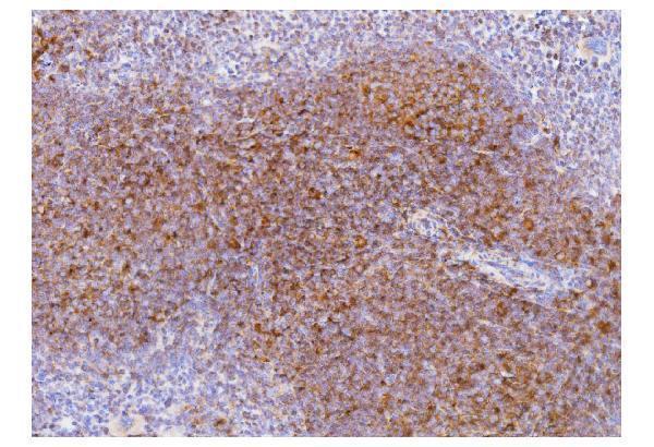
Click image to see more details
Figure 1. IHC analysis of Cd2 using anti-Cd2 antibody (A00570-1).
Cd2 was detected in paraffin-embedded section of mouse spleen tissue. Heat mediated antigen retrieval was performed in EDTA buffer (pH8.0, epitope retrieval solution). The tissue section was blocked with 10% goat serum. The tissue section was then incubated with 2μg/ml rabbit anti-Cd2 Antibody (A00570-1) overnight at 4°C. Biotinylated goat anti-rabbit IgG was used as secondary antibody and incubated for 30 minutes at 37°C. The tissue section was developed using Strepavidin-Biotin-Complex (SABC) (Catalog # SA1022) with DAB as the chromogen.

Click image to see more details
Figure 2. IHC analysis of Cd2 using anti-Cd2 antibody (A00570-1).
Cd2 was detected in paraffin-embedded section of rat spleen tissue. Heat mediated antigen retrieval was performed in EDTA buffer (pH8.0, epitope retrieval solution). The tissue section was blocked with 10% goat serum. The tissue section was then incubated with 2μg/ml rabbit anti-Cd2 Antibody (A00570-1) overnight at 4°C. Biotinylated goat anti-rabbit IgG was used as secondary antibody and incubated for 30 minutes at 37°C. The tissue section was developed using Strepavidin-Biotin-Complex (SABC) (Catalog # SA1022) with DAB as the chromogen.

Click image to see more details
Figure 3. IF analysis of Cd2 using anti-Cd2 antibody (A00570-1).
Cd2 was detected in paraffin-embedded section of mouse spleen tissue. Heat mediated antigen retrieval was performed in EDTA buffer (pH8.0, epitope retrieval solution). The tissue section was blocked with 10% goat serum. The tissue section was then incubated with 5μg/mL rabbit anti-Cd2 Antibody (A00570-1) overnight at 4°C. Biotin conjugated goat anti-rabbit IgG (BA1003) was used as secondary antibody and incubated for 30 minutes at 37°C. The tissue section was developed using DyLight®488 Conjugated Avidin (BA1128). The section was counterstained with DAPI. Visualize using a fluorescence microscope and filter sets appropriate for the label used.

Click image to see more details
Figure 4. IF analysis of Cd2 using anti-Cd2 antibody (A00570-1).
Cd2 was detected in paraffin-embedded section of rat spleen tissue. Heat mediated antigen retrieval was performed in EDTA buffer (pH8.0, epitope retrieval solution). The tissue section was blocked with 10% goat serum. The tissue section was then incubated with 5μg/mL rabbit anti-Cd2 Antibody (A00570-1) overnight at 4°C. Biotin conjugated goat anti-rabbit IgG (BA1003) was used as secondary antibody and incubated for 30 minutes at 37°C. The tissue section was developed using DyLight®488 Conjugated Avidin (BA1128). The section was counterstained with DAPI. Visualize using a fluorescence microscope and filter sets appropriate for the label used.
Protein Target Info & Infographic
Gene/Protein Information For CD2 (Source: Uniprot.org, NCBI)
Gene Name
CD2
Full Name
T-cell surface antigen CD2
Weight
39.448kDa
Alternative Names
CD2 antigen (p50), sheep red blood cell receptor; CD2 antigen; CD2 molecule; CD2; Erythrocyte receptor; FLJ46032; LFA-2; LFA-3 receptor; lymphocyte-function antigen-2; Rosette receptor; SRBC; T11; T-cell surface antigen CD2; T-cell surface antigen T11/Leu-5 CD2 LFA-2, SRBC, T11 CD2 molecule T-cell surface antigen CD2|CD2 antigen (p50), sheep red blood cell receptor|LFA-3 receptor|T-cell surface antigen T11/Leu-5|erythrocyte receptor|lymphocyte-function antigen-2|rosette receptor
*If product is indicated to react with multiple species, protein info is based on the gene entry specified above in "Species".For more info on CD2, check out the CD2 Infographic

We have 30,000+ of these available, one for each gene! Check them out.
In this infographic, you will see the following information for CD2: database IDs, superfamily, protein function, synonyms, molecular weight, chromosomal locations, tissues of expression, subcellular locations, post-translational modifications, and related diseases, research areas & pathways. If you want to see more information included, or would like to contribute to it and be acknowledged, please contact [email protected].
Specific Publications For Anti-Cd2 Antibody Picoband™ (A00570-1)
Hello CJ!
No publications found for A00570-1
*Do you have publications using this product? Share with us and receive a reward. Ask us for more details.
Recommended Resources
Here are featured tools and databases that you might find useful.
- Boster's Pathways Library
- Protein Databases
- Bioscience Research Protocol Resources
- Data Processing & Analysis Software
- Photo Editing Software
- Scientific Literature Resources
- Research Paper Management Tools
- Molecular Biology Software
- Primer Design Tools
- Bioinformatics Tools
- Phylogenetic Tree Analysis
Customer Reviews
Have you used Anti-Cd2 Antibody Picoband™?
Submit a review and receive an Amazon gift card.
- $30 for a review with an image
Be the first to review Anti-Cd2 Antibody Picoband™
*The first user to submit a review for a product is eligible for Boster's Innovating Scientists Reward, which gives product credits. This is in addition to the gift card reward.
Customer Q&As
Have a question?
Find answers in Q&As, reviews.
Can't find your answer?
Submit your question
